Human remains discovered and studied by archaeologists are in the majority of cases skeletal remains, with the exceptions being those that have been mummified, bodies preserved in bogs and rare cases of preservation in ice. This means that the primary means of analysing the demography and health status of past populations is restricted to studying the skeleton. Basic analysis of skeletal remains typically involves assessment of an individual’s age at death, biological sex (in adults), stature and pathology. These provide information on the life of individuals and when collated can provide insights into groups and larger populations. This section provides a summary of the basics of osteological analysis with key references and resources.
Terminology
The definition of childhood and the age at which someone becomes an adult are culturally defined and subject to considerable variation. As a result, bioarchaeologists refer to individuals with an estimated age of less than 17 years as ‘non-adults’ rather than children (Lewis 2007). The term ‘non-adult’ refers to any human skeletal remain or individual who has not reached biological or social maturity, generally encompassing newborns, infants, children, and adolescents up to about 17–18 years of age. Publications often contain the terms ‘infant’, ‘child’, ‘juvenile’ and ‘adolescent’ but the chronological ages referred to using these terms vary widely so care must be taken to correctly identify the age of the individuals under discussion.
Gender is also culturally defined and is distinct from biological sex. Bioarchaeologists estimate the biological sex of adult individuals on the basis of sexually dimorphic features on the skeleton. Individuals are categorised as male, female, or intermediate/indeterminable based on these features and the terms ‘male’ and ‘female’ refer strictly to biological sex and not to the individual’s gender.
Age and sex
Assessment of the age and sex of individuals is a vital component of understanding past populations and their health status. It is not always possible to accurately estimate the age and sex of individuals in past populations. This can be due to the variation in the rate of development and degeneration of an individual’s skeleton while they are living, as well as the variations in preservation of the skeleton following death, for example damage during burial rites such as cremation or due to taphonomic changes after burial.
Estimation of age is reliant on identifying stages of skeletal and dental development in non-adults and on the level of wear and degeneration in adults. Both skeletal development and degeneration vary between individuals and populations. Furthermore, skeletal development and degeneration can be impacted by external factors such as nutritional stress or type and degree of activity level. As a result, a variety of methods have been developed focusing on different aspects of development and degeneration for different populations.
Estimation of age in non-adults
In non-adults, age estimation is based on two methods: dental development and skeletal development and maturation.
Dental development follows a sequence of mineralisation and eruption that begins when the cusps of the deciduous (baby teeth) maxillary central incisors begin to mineralise at around 15 weeks of gestation and continues until the closure of the root apex of the permanent third molar at around 17 years of age (Lewis 2007, Schaefer et al 2009). The exact timing of this sequence varies between females and males and between populations but, on the whole, dental development is less affected by environmental factors and stress than skeletal development and is considered the most accurate method of estimating age in non-adults (Lewis 2007).
The development, growth and maturation of the skeleton begins with the initial formation of bones from around 5-6 weeks gestation and continues until skeletal maturation is largely complete at around 17 years of age. Estimation of age from this sequence of development takes the form of measurement of diaphyseal lengths (the shafts of the long bones such as the femur or humerus) and the appearance and fusion of epiphyses (ends of a long bone) to the diaphysis (Lewis 2007). Skeletal development is vulnerable to delays and disruption from factors such as nutritional stress or illness and there are notable variations in average timing of skeletal development between females and males and between populations. Data summarising age estimates based on skeletal development for a variety of populations is compiled in Juvenile Osteology (Schaefer et al 2009).
Estimation of age in adults
In adults, age estimation is based on the assessment of late fusing epiphyses, dental wear, changes to the auricular surface and pubic symphysis in the pelvis and changes to the sternal end of the fourth rib.
While the majority of skeletal development is complete at around the age of 17 years, there are a couple of late fusing epiphyses (for example the medial end of the clavicle and the iliac crest in the pelvis) which do not complete fusion until the individual is in their twenties (Schaefer et al 2009). The presence of epiphyses still undergoing fusion suggests an individual is in early adulthood.
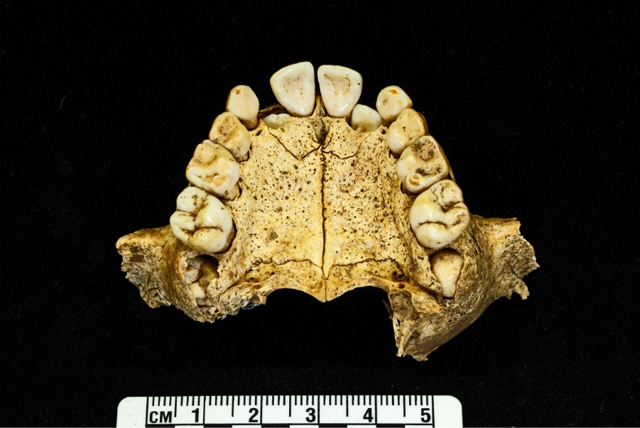
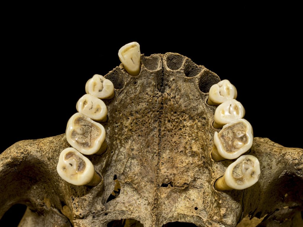
Adult age estimation from the dentition is based on examining the degree of wear of the teeth. The pattern and degree of dental wear can be influenced by dental disease and tooth loss but, provided these are taken into account, dental wear is considered a relatively reliable method of age estimation in adults. Brothwell’s (1981) method is frequently used in Britain for all populations up until the end of the medieval period, after which food typically becomes too soft for this method to be reliable.
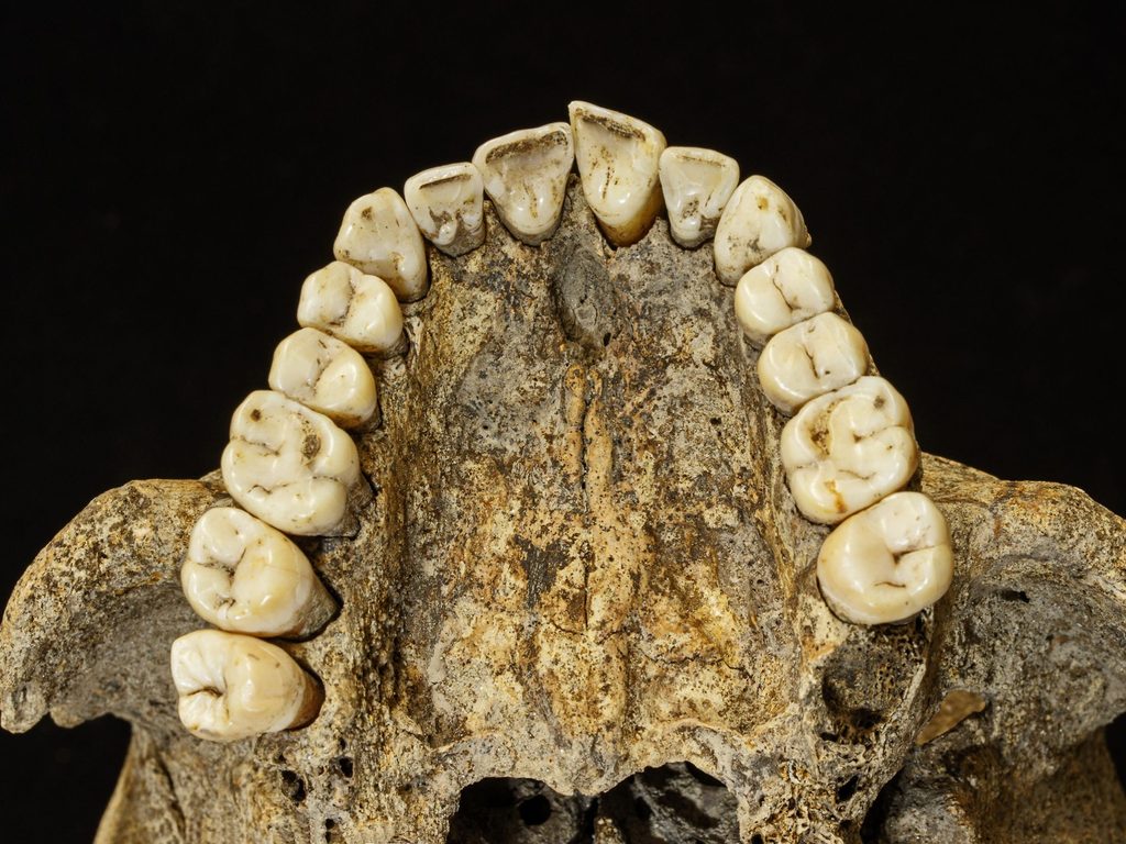
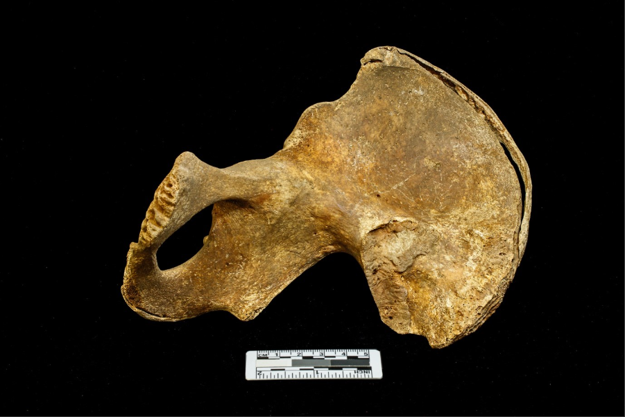
Estimation of adult age in the pelvis is based on age-related changes to the morphology and texture of the surface of the pubic symphysis and auricular surface. Estimation of age from the pubic symphysis entails examining changes to the surface following the descriptions laid out in Brooks and Suchey (1990). Estimation of age from the auricular surface also involves examining changes to the surface and comparison with the scoring system laid out in Buckberry and Chamberlain (2002). The auricular surface is more complex to score and less accurate than the pubic symphysis but it survives more frequently and does provide a broad age estimate.
Estimation of age from the sternal end of the fourth rib is based on changes to the morphology of the rib end and ossification of costal cartilage that progresses with age. The method proposed by İşcan et al (1984) divides the changes into 6 stages and provide age estimates into the fifth and sixth decades of life. However, its use is hampered by frequent difficulties in the preservation and correct identification of the fourth rib, separate standards being required for both race and sex and insufficient detail in the criteria resulting in inter-observer error (White et al 2012).
Assessment of sex
Assessment of biological sex in adults primarily relies upon sexually dimorphic features in the pelvis and skull. Many of these features are scored on a scale of 1-5, with a score of 1 indicating a typically female presentation of that feature and a score of 5 a typically male presentation, and some pelvic traits are assessed on the presence or absence of particular features. The range of scores and presence of features is then assessed to consider whether an individual appears to be female, male, or indeterminate. The age of the individual must also be considered as some post-menopausal females may develop more masculine cranial morphology and some young males have more gracile, feminine features. Diagrams and images of the key features are provided in Buikstra and Ubelaker (1994) and White (et al 2012).
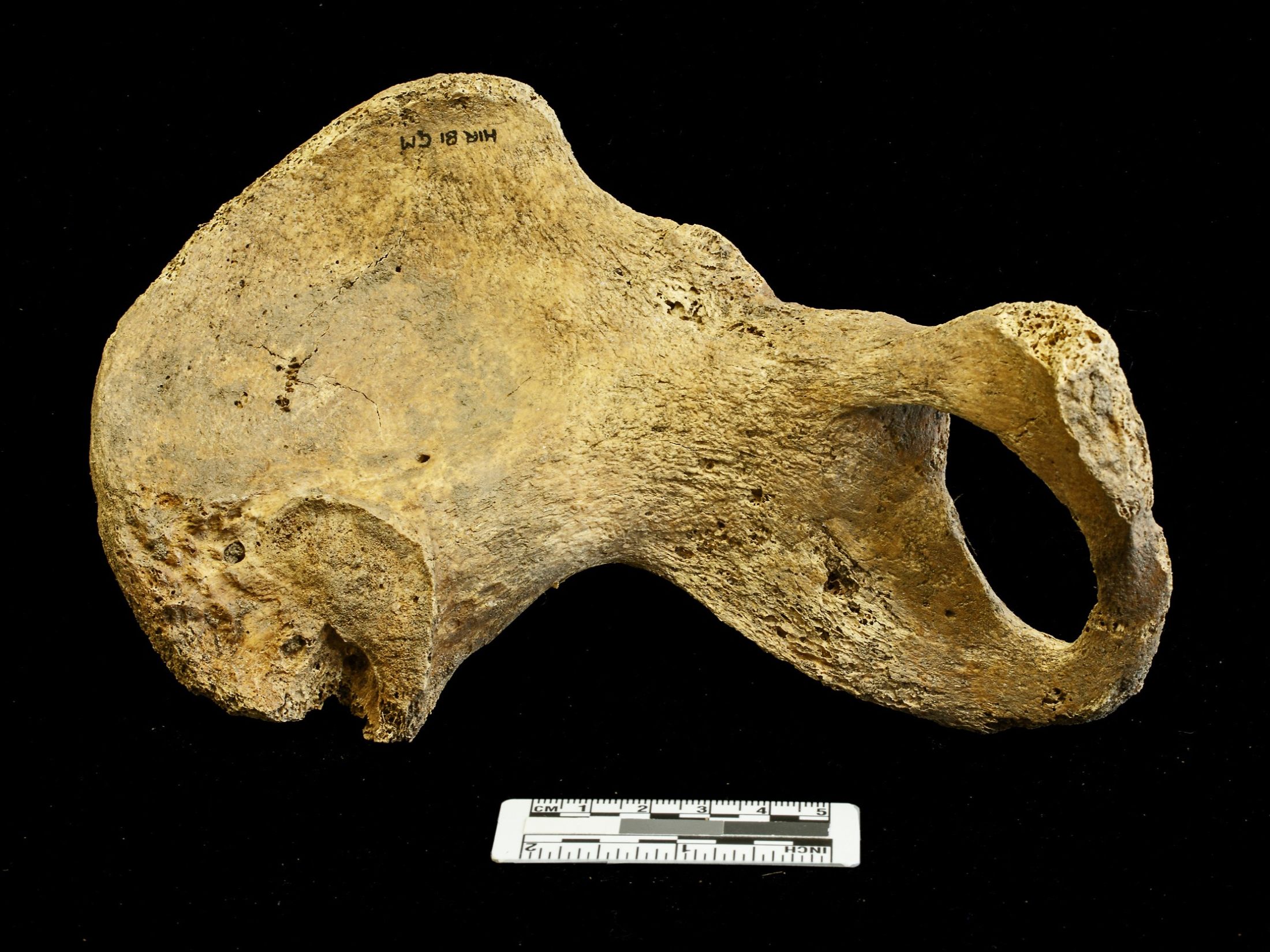
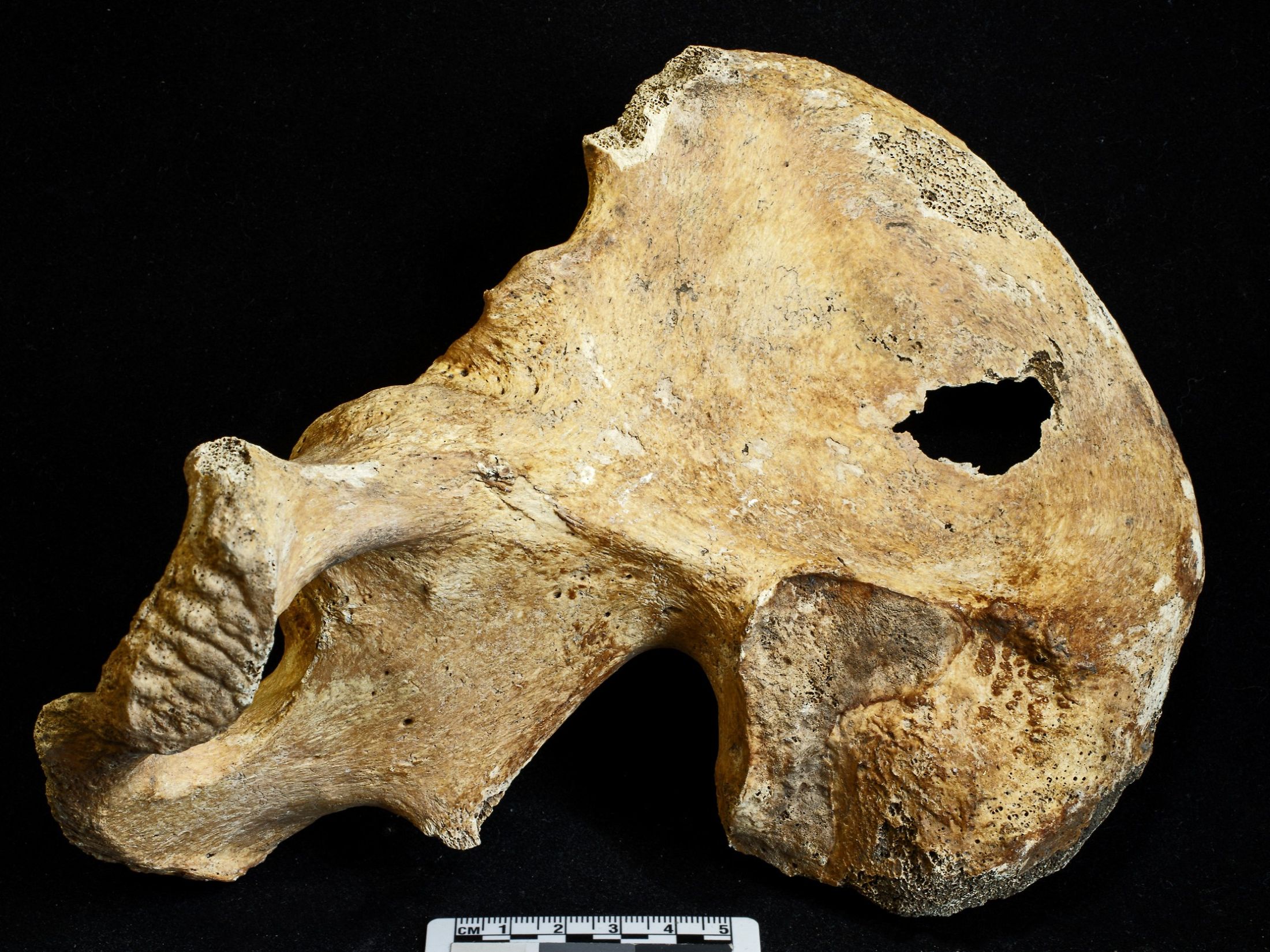
Metrical data can also assist in estimation of sex, especially in poorly preserved assemblages where the pelvis and skull may be damaged or missing. This method is less accurate as it relies on the average size of a male versus female bone and typical ranges of measurements vary between populations.
A great deal of research into the estimation of sex in non-adults has been undertaken, but as yet this work has not resulted in a method that is considered reliable. In recent years aDNA analysis of sex has become more common, but the destructive analysis and cost required means this is best used when answering a specific question where the biological sex of the individual is relevant.
Metrical analysis
In addition to assisting with estimation of sex, metrical data can also be used to estimate stature and examine other aspects of body size and shape. Full skeleton methods of estimating stature are the most accurate, but these are often difficult or impossible to use depending on quality of preservation or number of missing elements. Regression methods where the length of an individual bone is used in a regression-based equation remain the more common method of stature estimation. Many such equations exist but the equations derived from white and black American samples by Trotter and Gleeser (1977, 1958, 1952) are the most commonly utilised and these are summarised in Brickley and McKinley (2004).
Biocultural approach
In the past, skeletal analysis tended to concentrate on the individual. More recently, however, the thrust of osteological analysis is largely focussed on populations as seen within their cultural environment. While there may be variations in approach, the general emphasis today in osteology is to consider the ‘biocultural approach’. This term was first used in 1977 by Blakely:
‘Humans survive not through cultural adaptation nor through biological adaptation but through biocultural adaptation’.
Because culture is such an important component of human society, population groups must be understood within the context of their associated culture, which brings a far richer understanding of biological data.
The holistic biocultural approach has specific goals:
-
- To document specific ways in which biological anthropologists can contribute to studies of cultural processes.
- To illustrate the interrelationship between the biological, cultural and environmental variables that affect the adaptedness, or maladaptedness of prehistoric populations.
- To demonstrate the need for co-operation among biological anthropologists, archaeologists, ethnologists and other expert investigators toward problem-solving in behavioural anthropology. The biocultural approach has led to a number of interdisciplinary studies, including:
-
- Bone chemistry for dietary reconstruction
- Palaeodemographic studies
- Palaeoepidemiology
- Field anthropology
- Biological distance studies
- Biomechanical approaches for understanding patterns of activity
- Stable isotope ratios (strontium) for establishing mobility.
-