In addition to the standard macroscopic methods of analysis there are a range of analytical methods that can provide further insight into skeletal assemblages. These include imaging techniques (eg radiographs), destructive analyses (such as stable isotope analysis or radiocarbon dating), and associated areas of study (such as palaeoparasitology). This section summarises the application of these techniques to archaeological human remains but full updated sections for commonly applied techniques are to be developed in due course.
Imaging Techniques
Imaging techniques are used to assist in the visualisation or diagnosis of pathology (Villa et al 2019). Radiographs are used to view some pathological lesions, evidence of growth stunting and evidence of trauma that may not be visible on the surface of the bone. For example, well-healed trauma can sometimes be difficult to identify but will appear as an area of increased density on an X-ray. Computed tomography (CT scans) and magnetic resonance imaging (MRI scans) are also used to further analyse pathology that is hidden within the shaft of a bone or on the interior of an intact skull.
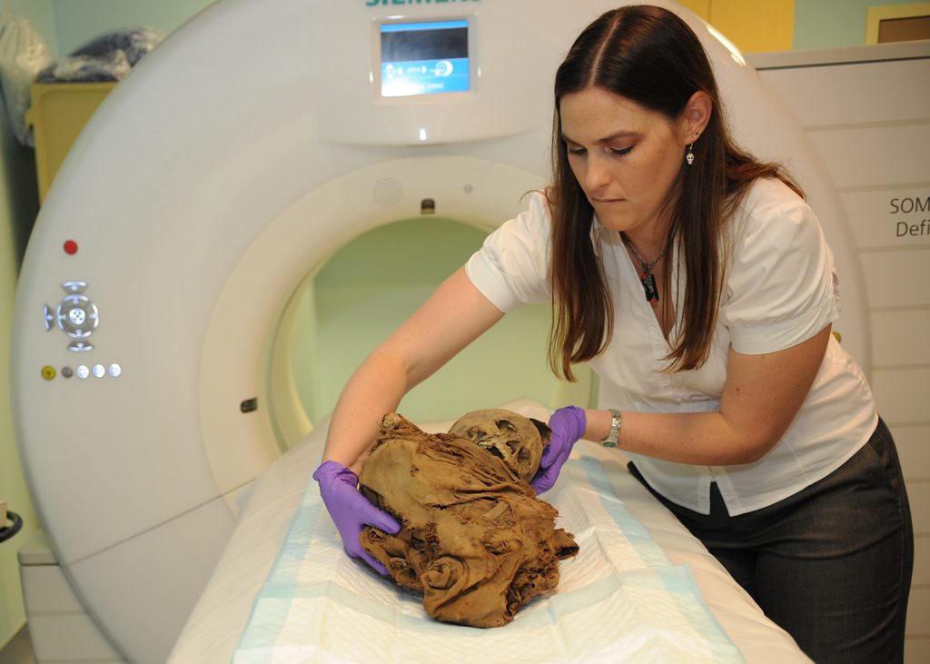
CT scans and MRIs can be used to create 3D models of bone, which can also be created through laser or light scanning and photogrammetry. 3D modelling is used in a variety of ways, including creating the base model for facial reconstructions and investigating differences in skeletal morphology between populations or changes over time (Gunz 2019, Maclennan et al 2025, Wilkinson et al 2023).
Destructive Analyses
Radiocarbon dating, stable isotope analysis and aDNA (ancient DNA) analysis all require samples of bone or tooth to be taken, which are destroyed during the analysis process. As such, samples should be as small as possible and well documented with photographs or photogrammetry. They should also be taken from areas of bone or tooth that are not affected by pathology, trauma, or used in other techniques such as metrical analysis.
Use of these techniques has increased in recent years due to both major advancements in each technique and a fall in the cost of analysis. Positive results can now be obtained from relatively small samples of bone and when combined with falling costs this has made these techniques increasingly accessible and applicable to a wider range of productive research avenues (Becerra-Valdivia and Higham 2023, Orlando et al 2021, Stantis et al 2025).
Radiocarbon dating of human remains is used both to identify the date of the remains themselves and to date associated sites and artefacts. In the case of cemetery sites that were in use for an extended period, radiocarbon dating of multiple individuals can be used to investigate the period of use and phases of activity. Human remains sampled for aDNA research are usually also radiocarbon dated. Any dates obtained from material within the collections of the National Museums of Scotland are published annually in Discovery and Excavation in Scotland, including dates procured as part of large aDNA projects (Knight et al 2023).
Stable isotope analysis can be used to explore migration and dietary trends in past populations. The primary isotopes used are carbon, nitrogen, oxygen and strontium, although data on sulphur isotopes is increasing. Oxygen, strontium and sulphur can provide information about migration of individuals, while carbon and nitrogen provide information about diet in relation to the types of plants and animal or marine proteins individuals were eating.
Teeth do not remodel once formed, but the skeleton continues to remodel and turn over fresh bone throughout life. Isotopic analysis of different elements can provide information on where an individual lived and what they ate at different phases of life (see Case Study: Dietary Isotope Analysis at Portmahomack). Isotopic analysis of both teeth and bone from one individual can highlight whether they spent their childhood in a different area (Müldner et al 2009). Incremental sampling of dentine from inside teeth can provide insights into weaning practices as the introduction of food and the cessation of breastfeeding alters nitrogen isotope values (Britton et al 2018).
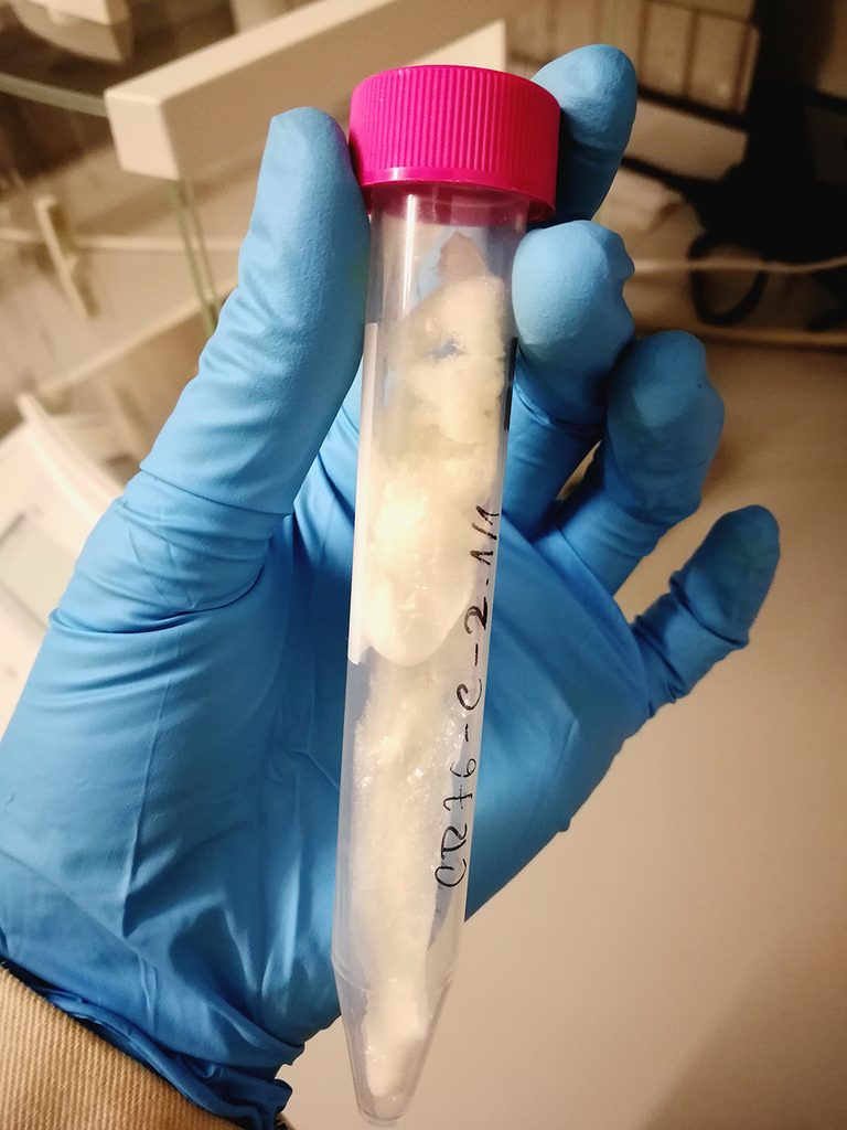
Ancient DNA analysis is a comparatively young field but rapid advancements have led to an explosion of aDNA research in recent years. aDNA analysis of bone has been applied to ancient genomics and exploring the origins of specific genetic variations. Samples from the pulp cavity of teeth and dental calculus have also been analysed to investigate the genetic development of pathogens such as tuberculosis and Yersinia pestis and, in the case of calculus, examine the history of the oral microbiome (Gancz et al 2023, Schuenemann et al 2011).
Further Associated Techniques
Palaeoparasitology is the study of ancient parasites. In bioarchaeological terms this is mostly done through analysis of samples from latrines or from the abdominal area of graves. Parasites are largely identified by their eggs and the presence of particular species can provide insights into health status and sanitation practices (Mitchell 2015).
Zooarchaeology can provide insights into past populations as well as the animals they interacted with (Upex and Dobney 2020). Animal bones can provide information on what a group were eating, and the zoonoses (infections passed from animals to humans) that they might have been exposed to.
Proteomics is the study of ancient proteins and focuses on identifying proteins, or their components, through mass spectrometry (Hendy et al 2019). This has been used to identify species in fragmentary comingled deposits of bone (Buckley and Kansa 2011) and to identify dietary proteins preserved in dental calculus (Warinner et al 2014a).
Dental calculus has become an interesting focus of study in recent years through the analysis of proteins, oral biome and pathogen aDNA, as well as microfossils and debris trapped within its matrix. Proteins and aDNA have shown the history of the oral microbiome, including identifying bacteria strongly associated with periodontal disease (Warinner et al 2014b). Microfossils and fragments of environmental debris have provided insights into the diets and even occupations of past individuals (Fagernäs and Warinner 2023, Radini et al 2019).
X-ray fluorescence spectrometry (XRF) is a non-destructive method of analysing chemical composition that has a wide range of applications in archaeology. It is frequently used in analysis of lithics, glass and metal artefacts, as well as soil mapping, but also has applications to human remains (Shackley 2018, Williams et al 2020). XRF analysis of the composition of bones has been used to investigate diagenetic changes to the bone, separate commingled remains of multiple individuals and examine burning temperature in cremated remains (Gomes et al 2024, McGarry et al 2021).