Palaeopathology
Palaeopathology is the scientific study of ancient diseases, injuries, and disorders in humans, animals, and plants through the examination of skeletal remains, mummified tissues and fossils.
Identifying and accurately recording pathological lesions in human skeletal remains is a vital component of the analysis and understanding of past populations. However, there are inherent limitations to recording pathology in skeletal remains, primarily that the pathology observed in the skeleton provides only a partial view into an individual or group. Many diseases do not impact bone or only do so in a few cases and acute infectious diseases often leave no trace. With the exception of perimortem trauma (trauma occurring around the time of death), it is usually impossible to identify a definite cause of death from the skeleton alone.
The key component of analysing skeletal pathology is to accurately describe the type, location and extent of any bony changes observed. The aim is to describe all pathology present so that other researchers can understand what was observed, rather than making a diagnosis for each individual lesion. This is often followed by a differential diagnosis which describes the most likely diagnosis and other potential causes.
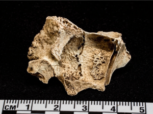
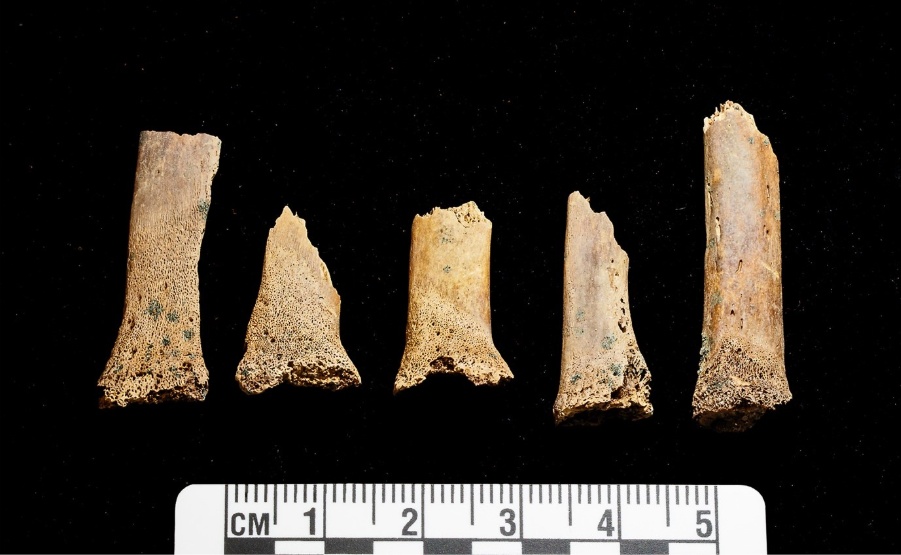
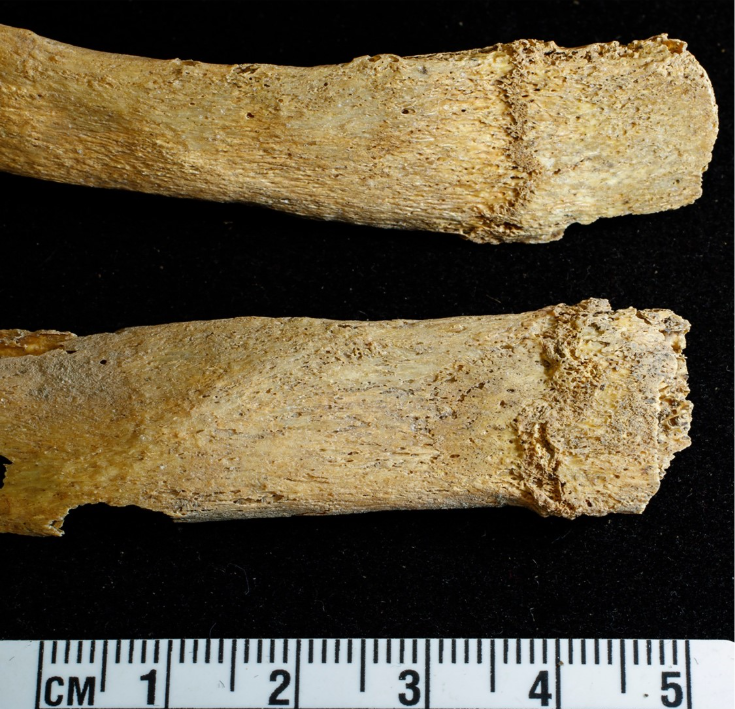
The example below shows the flared and cupped sternal ends of the ribs of an individual aged 1-2 years from Constitution Street, Edinburgh. This is suggestive of either rickets (vitamin D deficiency) or scurvy (vitamin C deficiency), and the diagnosis would depend on the other pathology present. In bioarchaeology, these lesions are more consistently associated with vitamin D deficiency but have been observed in individuals with scurvy both in archaeological human remains and in living children in clinical medicine (Brickley and Morgan 2023, Snoddy et al 2018).
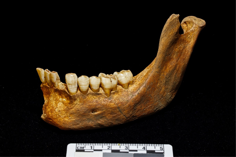
Some pathological lesions are caused by a variety of stressors and cannot identify a specific illness or type of stress. Instead, these serve as an indicator that the individual had experienced a significant episode or period of stress. Linear Enamel Hypoplasias (LEH) are horizontal defects in tooth enamel that indicate that a stressor, such as illness or nutritional stress, interrupted the normal development of the tooth crown (Hillson 2014). Since tooth enamel does not remodel once grown, these defects are permanent. LEH is commonly examined as an indicator of periods of stress experienced during childhood (Armelagos et al 2009, Miszkiewicz 2015).
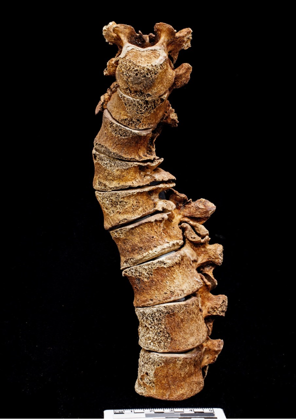
The identification of pathology is not restricted to lesions, and also includes changes in normal bone development, shape or robusticity that indicate congenital and developmental abnormalities and evidence of limb disuse (Lewis 2019a, 2019b). Identifying severe genetic disorders and congenital abnormalities is rare in archaeological contexts. However, other disorders, including cleft palate and spina bifida, are more commonly observed. Developmental abnormalities, such as scoliosis, can also be identified, as shown in the example below of a medieval individual from Constitution Street in Edinburgh. Long term limb disuse can result in underdevelopment or wasting of the limb in question and is identified through a difference in size and robusticity between the paralysed and functional limbs. This can be indicative of poliomyelitis infection, stroke, neurological disorders or nerve injuries (Berner et al 2021, Grauer and Roberts 2019).
Additional analysis may be required to identify some pathology. Osteoporosis, for example, is only macroscopically visible through the presence of fractures that commonly occur in individuals with low bone density. However, those fractures may not occur in every case and also still occur in individuals without osteoporosis. Therefore, accurate assessment of osteoporosis requires the use of X-ray absorptiometry to measure bone density or radiographs to measure thinning of cortical bone (Brickley and Mays 2019). Well-healed trauma, evidence of growth interruption (ie Harris lines), and some cancers are often only observed through the use of radiographs or CT scans. Recent work with dental histology samples has found correlation between interglobular dentine and vitamin D deficiency during development, though more work is needed to establish a threshold for diagnosis of deficiency (Snoddy et al 2024).
There are some circumstances in which it is possible to identify the likely presence of a disease that does not produce skeletal pathology. In some cases, outbreaks of infectious disease can be identified through the presence of mass graves or burial grounds in use for an extremely short period of time.
The largest known example of this in Britain is the assemblage of 636 individuals from the East Smithfield Black Death cemetery in London, which was in use for a period of two years during the major outbreak of Yersinia pestis in AD 1348-1350. Analysis of aDNA fragments in tooth and bone samples confirmed the presence of Y. pestis in these individuals (Schuenemann et al 2011).
The use of aDNA to search for evidence of infectious diseases has been increasing in recent years. In addition to providing evidence of Y. pestis being the pathogen responsible for the Black Death, analysis of pathogen aDNA has provided insights into the history of tuberculosis and leprosy and how those pathogens have changed over time (Donoghue 2019, Donoghue et al 2015, Kerner et al 2021, Murphy et al 2026).
The presence of parasites can also be identified through analysis of soil samples from latrines and the abdominal area of graves for parasite eggs, as well as the remains of the parasites themselves. A variety of gastrointestinal parasites have been identified in British archaeological sites (Mitchell 2015), including roundworm and whipworm eggs found in a latrine in medieval Perth (Holdsworth 1987), which provides insights into both sanitation practices and health.
Limitations of bone and the Osteological paradox
Studying archaeological human remains to infer the health or cause of death of individuals requires engaging with the inherent limitations of the skeleton. Many diseases do not impact bone, or only do so in a handful of cases, and even when pathology is present it is rare to be able to identify a potential cause of death. Bone has a limited number of responses to stimuli and changes primarily occur through the action of blastic (bone forming) or clastic (bone destroying) cells. The balance and pattern of bone formation versus destruction can be indicative of a particular cause but often the changes are non-specific or can only indicate a general category of stressor or disease process. For example, periosteal new bone formation can occur in response to infections, repetitive strain injuries (eg shin splints) and some nutritional deficiencies.
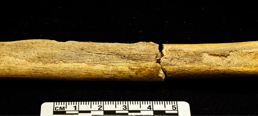
In addition to the limited response of bone, changes to bone take more time to develop than changes to soft tissue, meaning that many pathological lesions in the skeleton have required an individual to survive for days, weeks or longer in order for that lesion to develop.
The time required for skeletal pathology to develop also means that for many diseases, the frailest individuals may not survive for long enough to show any bony changes. As a result, some individuals with significant skeletal pathology may in fact be some of the more resilient individuals. This is known as the Osteological Paradox, which must be considered when interpreting health in past populations (Wood et al 1992).
Taphonomy
Taphonomy is the study of what happens to organic remains after death. Understanding the impact of these ‘taphonomic processes’ on human remains is paramount to bioarchaeological analysis. Chemical and microbial degradation, plant root and fungi action, and gnawing or burrowing from rodents and insects can all take their toll on the preservation of bone. The hydrology of a site also has an impact, with repeated cycles of wetting and drying tending to result in much poorer preservation than in waterlogged sites (Pinhasi and Mays 2008).
The impact of taphonomic processes can vary throughout the skeleton. Typical bone density varies across different skeletal elements with the major bones of the cranium and the shafts of the long bones containing more dense cortical bone while the vertebrae, pelvis and other elements contain more trabecular bone (White et al 2012). Dense cortical bone which is less likely to be damaged within the grave, survives better in poor soil conditions and is more likely to survive in identifiable fragments in cremated remains.
The open, spongy texture of trabecular bone means that it is more vulnerable to degradation and damage, biasing elements with a high proportion of trabecular bone towards poor preservation. Dental enamel is the hardest material in the human body (Lynnerup and Klaus 2019) and as a result teeth can often survive better than the surrounding bone.


The bones of non-adults are less well mineralised than those of adults and more vulnerable to taphonomic damage. The small size and relative fragility of the skeletons of infants and younger children increases the chance of fragmentation and further degradation, while the skeletons of adolescents show a pattern of preservation more similar to adults (Lewis 2007).
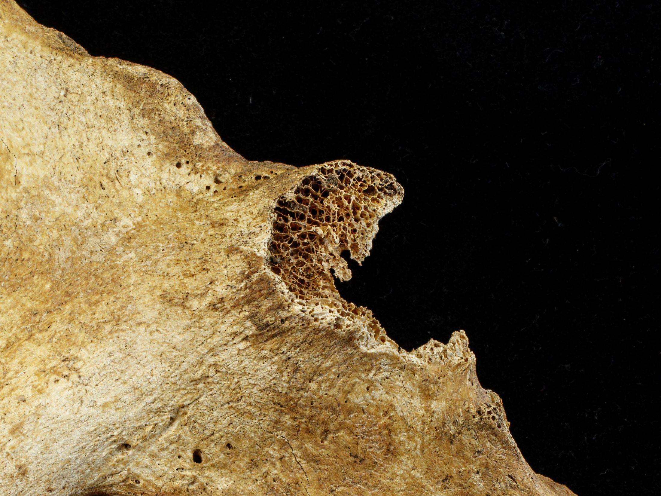
Some pathological changes are vulnerable to taphonomic damage. The fine spicules of new bone formation that occur on the bone surface or at the edge of an injury are delicate and vulnerable to be damaged or obliterated if preservation is poor. Pathological lesions that involve destruction of bone can also be damaged beyond identification or bias a particular bone or individual towards poor preservation (Brickley and Mays 2019, Marques 2019).
Damage to bone during burial, due to taphonomic processes and during excavation, can also result in pseudopathology or alterations to the bone that mimic dieseases or a medical condition. This includes post-mortem fractures, damage from the growth of salt crystals and ‘trowel trauma’ (Klaus and Lynnerup 2019).
The circumstances of a site and how soil conditions and post-deposition disturbance may have impacted an assemblage must be taken into account during analysis.