X-ray Fluorescence (XRF)
Energy-dispersive XRF
This technique uses an X-ray beam to excite fluorescent X-rays from the surface of a sample. These are detected by a solid state detector (usually liquid nitrogen-cooled Si(Li), although room temperature detectors are becoming more common) which measures their energy and quantity. Elements are identified from their unique set of fluorescent X-ray energies and composition can be deduced from their relative abundance in the XRF spectrum under the experimental condition.
XRF has the advantage of being completely non-destructive and very quick. An air-path system can be used directly on complete objects.
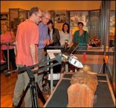
pXRF of Egyptian sarcophagus 1895 167a, Kelvingrove Museum, Glasgow (photo: Glasgow Museums)
The main limitation of the technique is that it is only able to analyse the surface layer (typically up to the first hundred microns, but the depth depends on the material composition). This means that if surface enrichment has taken place, for example on a metal artefact, the results may not represent initial bulk composition (Tate 1986). XRF can only detect light elements such as sodium if the sample is maintained in a vacuum chamber, a system with reduced suitability for much archaeological material or complete artefacts.
XRF has been used to characterise archaeological metals, such as copper alloys and silver, and is also highly suited as first-line identification technique for a wide range of other inorganic materials and to characterise surface gilding or decoration, Micro-XRF systems are now well established where the analysed area is of micron dimensions and may be used to build up a compositional maps across small areas of an artefact’s surface.
Used at: NMS, University of St Andrews (See also the ScARF Case Study on the Duart Point shipwreck in the Marine and Maritime panel report)
p-XRF

Figure 14: 16thc Asian bronze buddah (Glasgow Museums 1929.64.ca)
Portable or hand-held X-ray fluorescence has been used intermittently in archaeological science for the last couple of decades, however recent advances in detector technology, specifically the introduction of the silicon drift detector (SDD) means that it is becoming more popular not only across a range of material types but also as an ideal in-situ ‘screening’ technique, for example on artefacts in museums. Various techniques (partial vacuum, He flush) allow systems to detect elements as light as Al.
Used at: HS, Scottish Analytical Services for Art & Archaeology (Glasgow)
A different method of determining the energy of the fluorescent X-rays (see XRD below) can lead to higher sensitivity. The method is often used for bulk sample analysis of stone, soil and ceramics. Detects a wide range of elements, and as the sample is normally contained in a vacuum chamber, includes light elements.
Used at: Edinburgh Geosciences,
X-ray diffraction (XRD)
X-ray diffraction provides information about the structure of a material, rather than its chemical composition. X-ray diffraction depends on the X-ray wavelength and the lattice spacing of the sample material, by measuring the angles at which diffraction lines occur the crystalline structure of the sample can be deduced. This technique is used to identify minerals and geological materials, to characterise corrosion products of metals and deterioration products of stone and in any aspect of archaeological science where knowing the structure as well the composition of a material is required.
Used at: Edinburgh Geosciences, Historic Scotland, NMS, Glasgow Earth Sciences, Charleston Consultants (Scottish Lime Centre, Fife, University of St Andrews
Electron Microprobe analysis (EPMA) and Scanning Electron Microscopy (SEM) with Energy or Wavelength Dispersive analysis (EDS or WDS)
In each of these methods the sample, normally as a small polished specimen mounted in a plastic block and carbon coated, is irradiated with a beam of electrons. Fluorescent X–rays are generated by the electron-sample interaction and can be detected as in XRF, using energy dispersive or wavelength dispersive spectroscopy. The electron microprobe uses a highly focused and intense electron beam to analyse all but the lightest elements (>Be) with a spatial resolution of about 1micron and good analytical precision. The scanning electron microscope (see below) allows similar analysis but with lower sensitivity in favour of improved image resolution. EPMA has been applied mostly to glass, whereas SEM-EDS is commonly used for a wide range of metals and vitreous materials.
Used at: NMS (SEM-EDS) Edinburgh Geosciences ((EMPA/SEM-WDS) where it forms part of the NERC Tephrochronology Service)
Particle induced X-ray emission (PIXE) and Gamma spectroscopy (PIGE)
This technique uses a finely focused ion beam, normally protons, to excite X-rays from the sample. Like XRF, the energy of these X-rays is diagnostic of each element present. PIXE allows quantitative analysis with very low detection limits for many elements (ppm) and has been applied to the trace element characterisation of metals, ceramics and lithics. The proton interaction also causes gamma emission from some elements: this emission is not just from the surface of the sample and thus gamma spectroscopy can give information about the bulk, as well as surface composition of a sample. Various other techniques such as Rutherford backscattering analysis can be combined with this methodology.
Used at: Department of Materials at Oxford, CNRS Louvre Paris
Analysis using Synchrotron radiation
At the high-tech end of analytical methods is the national synchrotron science facility, Diamond Light Source, based at Harwell. The unique characteristics of synchrotron radiation – particularly the ability to produce a high intensity and tightly focused excitation beam of electromagnetic radiation – are ideal for analytical problems requiring high spatial or temporal resolution or problems that are intractable using conventional instruments. Cultural heritage work exploits analytical techniques from infrared to hard X-ray wavelengths. Imaging from X-ray to micro-CT scanning is also possible.
Fourier Transform Infra-red Spectroscopy (FTIR)
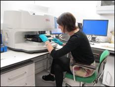
Figure 15: FTIR microscope in use at NMS Collection Centre, Edinburgh ©NMS
This technique identifies materials based on their absorption of infra-red light. The absorption spectrum depends on the molecular species present since they have different vibrational frequencies. It is most commonly used in the identification of organic materials such as waxes, resins and pigments, which, in a conservation context, are helpful in determining previous treatments on objects. Modern materials including plastics can also be routinely identified with FTIR. FTIR can be used in bench mode or with various microscope configurations for the examination of micro-samples. Interest is now focused on hand-held instruments for in-situ analysis.
Used at: NMS, Textile Conservation Centre Glasgow, Strathclyde, Napier (Forensics)
Raman spectroscopy, Fourier Transform Raman spectroscopy, Surface Enhanced Raman Spectroscopy.
Like FTIR, but using a visible laser rather than infra-red, Raman spectroscopy provides information about the molecular species present in a sample and their charge state. This technique has been extensively applied to a wide range of materials. It has been applied routinely to pigment identification and has also been used to look at the corrosion of glass and other materials in a museum setting.. In materials which have transparent upper layers the area beneath can be analyses, allowing examination of, for example, pigment layers beneath glass, while significant improvements in detection limits can be obtained with SIMS. In recent years the use of surface enhanced Raman scattering (SERS) has greatly enhanced the capabilities of Raman as a technique, significantly reducing the size of sample required.
Used at: Glasgow Earth Sciences (Renishaw inVia), Strathclyde, Napier